

AI-ACCELERATED DRUG DISCOVERY
Available from Reaxense
This protein is integrated into the Receptor.AI ecosystem as a prospective target with high therapeutic potential. We performed a comprehensive characterization of Epithelial cell adhesion molecule including:
1. LLM-powered literature research
Our custom-tailored LLM extracted and formalized all relevant information about the protein from a large set of structured and unstructured data sources and stored it in the form of a Knowledge Graph. This comprehensive analysis allowed us to gain insight into Epithelial cell adhesion molecule therapeutic significance, existing small molecule ligands, relevant off-targets, and protein-protein interactions.
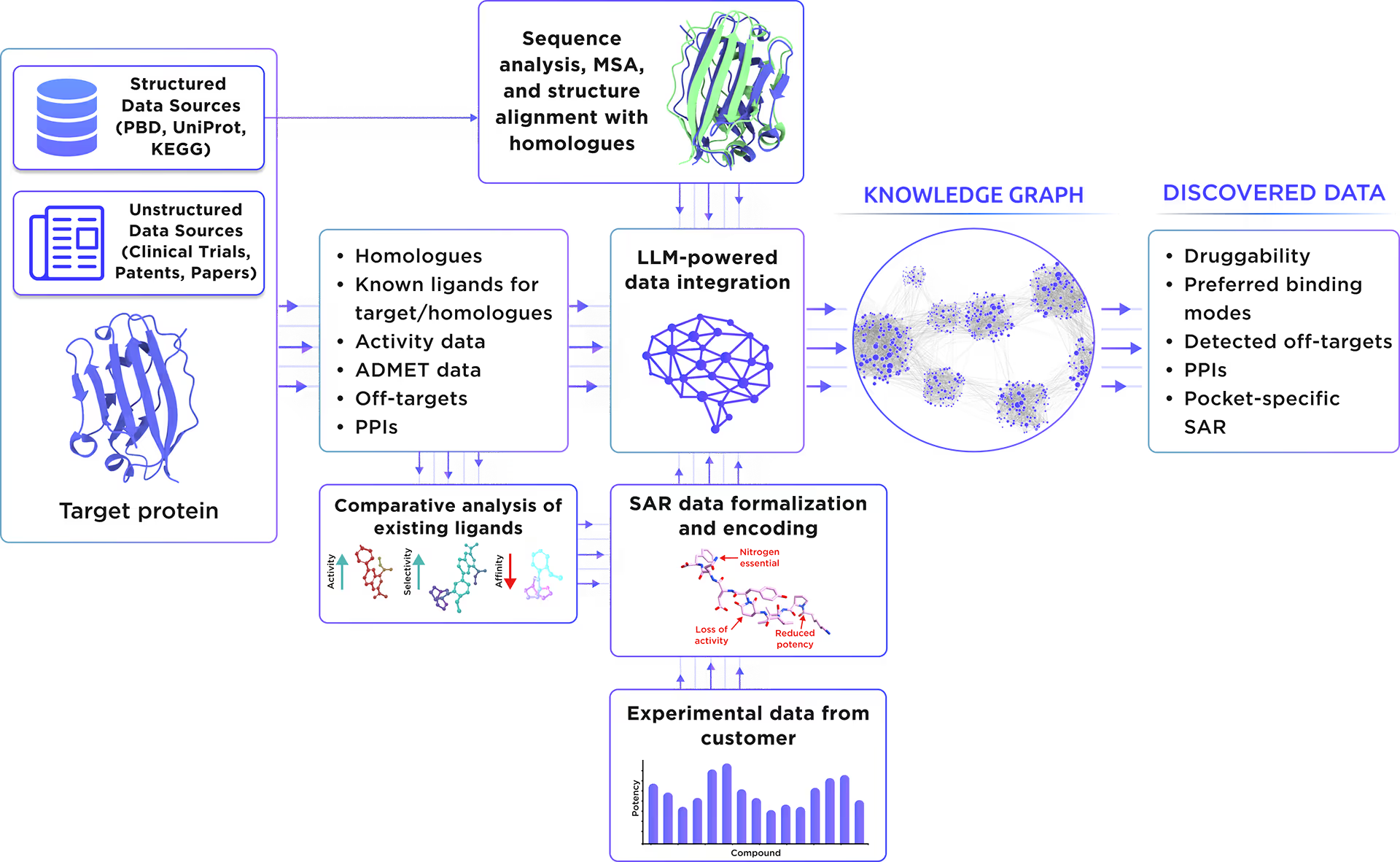
Fig. 1. Preliminary target research workflow
2. AI-Driven Conformational Ensemble Generation
Starting from the initial protein structure, we employed advanced AI algorithms to predict alternative functional states of Epithelial cell adhesion molecule, including large-scale conformational changes along "soft" collective coordinates. Through molecular simulations with AI-enhanced sampling and trajectory clustering, we explored the broad conformational space of the protein and identified its representative structures. Utilizing diffusion-based AI models and active learning AutoML, we generated a statistically robust ensemble of equilibrium protein conformations that capture the receptor's full dynamic behavior, providing a robust foundation for accurate structure-based drug design.
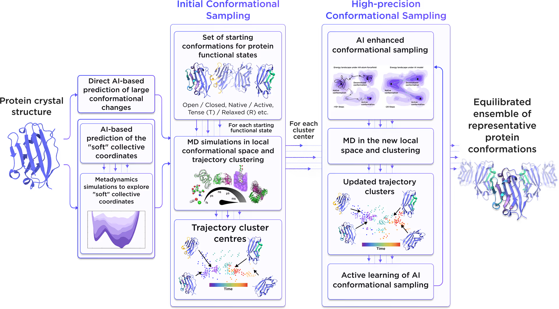
Fig. 2. AI-powered molecular dynamics simulations workflow
3. Binding pockets identification and characterization
We employed the AI-based pocket prediction module to discover orthosteric, allosteric, hidden, and cryptic binding pockets on the protein’s surface. Our technique integrates the LLM-driven literature search and structure-aware ensemble-based pocket detection algorithm that utilizes previously established protein dynamics. Tentative pockets are then subject to AI scoring and ranking with simultaneous detection of false positives. In the final step, the AI model assesses the druggability of each pocket enabling a comprehensive selection of the most promising pockets for further targeting.
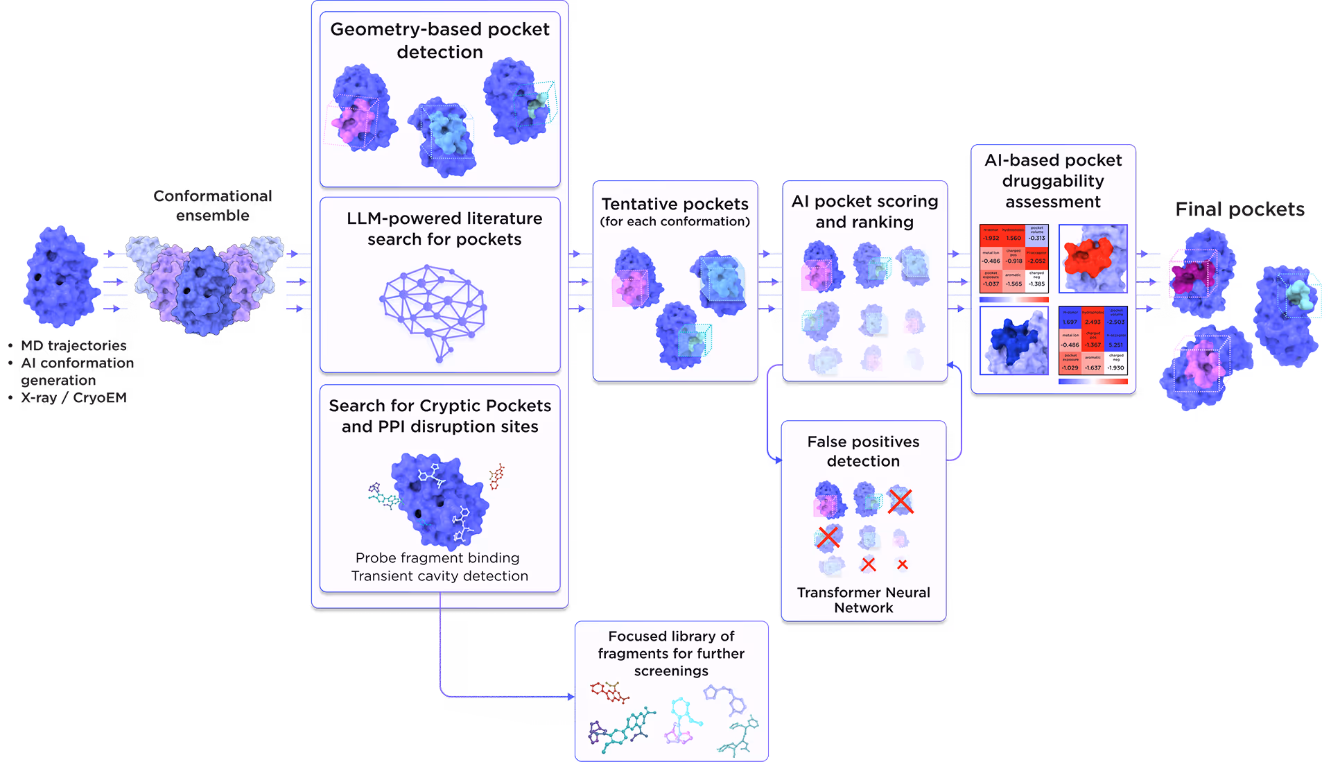
Fig. 3. AI-based binding pocket detection workflow
4. AI-Powered Virtual Screening
Our ecosystem is equipped to perform AI-driven virtual screening on Epithelial cell adhesion molecule. With access to a vast chemical space and cutting-edge AI docking algorithms, we can rapidly and reliably predict the most promising, novel, diverse, potent, and safe small molecule ligands of Epithelial cell adhesion molecule. This approach allows us to achieve an excellent hit rate and to identify compounds ready for advanced lead discovery and optimization.
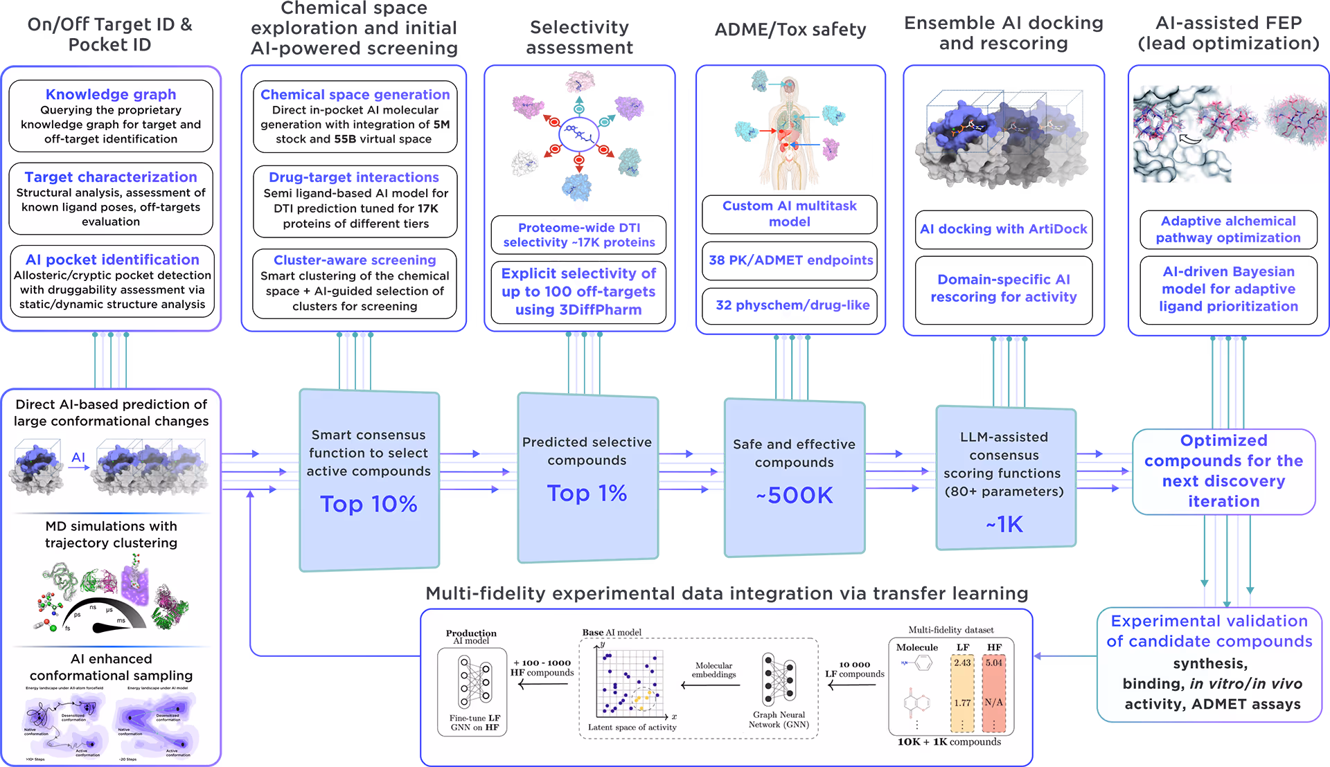
Fig. 4. The screening workflow of Receptor.AI
Receptor.AI, in partnership with Reaxense, developed a next-generation technology for on-demand focused library design to enable extensive target exploration.
The focused library for Epithelial cell adhesion molecule includes a list of the most effective modulators, each annotated with 38 ADME-Tox and 32 physicochemical and drug-likeness parameters. Furthermore, each compound is shown with its optimal docking poses, affinity scores, and activity scores, offering a detailed summary.
Epithelial cell adhesion molecule
partner:
Reaxense
upacc:
P16422
UPID:
EPCAM_HUMAN
Alternative names:
Adenocarcinoma-associated antigen; Cell surface glycoprotein Trop-1; Epithelial cell surface antigen; Epithelial glycoprotein; Epithelial glycoprotein 314; KS 1/4 antigen; KSA; Major gastrointestinal tumor-associated protein GA733-2; Tumor-associated calcium signal transducer 1
Alternative UPACC:
P16422; P18180; Q6FG26; Q6FG49; Q96C47; Q9UCD0
Background:
The Epithelial Cell Adhesion Molecule (EpCAM), also known as Tumor-associated calcium signal transducer 1, plays a crucial role in cell-cell adhesion, providing an immunological barrier against mucosal infection. It is involved in the proliferation and differentiation of embryonic stem cells and up-regulates the expression of key proteins such as FABP5, MYC, and cyclins A and E.
Therapeutic significance:
EpCAM's association with Diarrhea 5, characterized by intestinal epithelial cell dysplasia, and Lynch syndrome 8, linked to increased cancer susceptibility, underscores its potential as a target for therapeutic intervention in both congenital intestinal disorders and hereditary cancers.